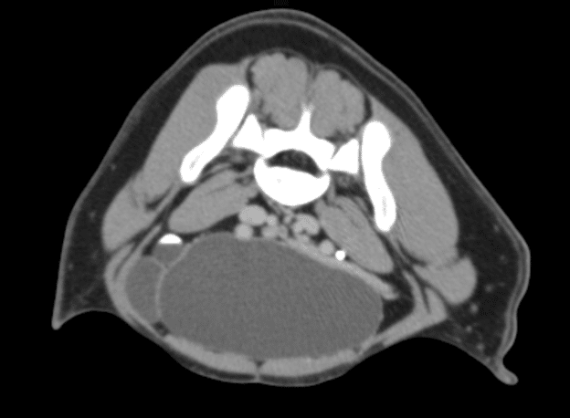
Canine Ultrasound & CT Case Study

Wheatie (Rye) is a 6-month-old intact female Vizsla who first presented to her referring veterinarian for recurrent urinary tract infections treated with repeated antibiotic therapy. As a board-certified internal medicine specialist, the veterinarian performed a complete diagnostic evaluation to identify a cause for the recurrent infections including bloodwork, urinalysis, abdominal radiographs, and an abdominal ultrasound.
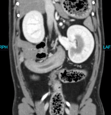
Canine Scintigraphy and CT

Copper is a 4 year female old Shih Tzu who had a history of elevated liver enzymes. Copper’s ALT was 700 (rr 0-120), pre bile acids were 84.7 (rr <13), and post bile acids were 256.4 (rr <25). With these lab values, the referring veterinarian was concerned for a possible shunt and Copper was referred to Animal Imaging.

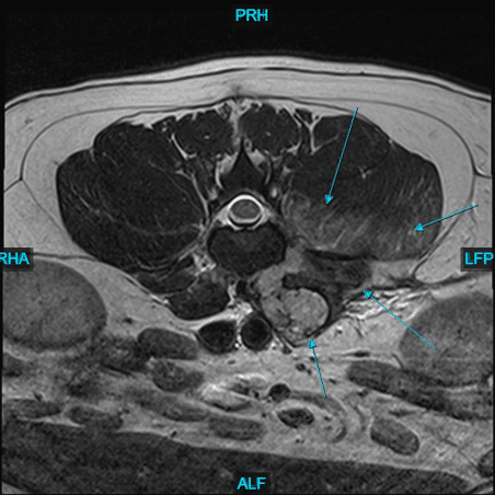
Canine Orthopedic Advanced Imaging

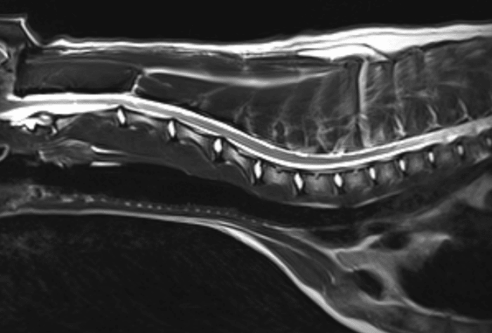
Benji, a 1-year-old intact male Golden Retriever, presented to Animal Imaging for advanced imaging of his right shoulder, brachial plexus, and elbow. He had a 9-month history of right front limb lameness that was worse after prolonged periods of laying down or intense exercise.

Equine MRI Suspensory Study

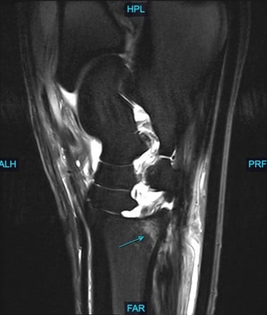
Hugo is a 17 year old Warmblood gelding used for show jumping. Hugo had a 12 month history of acute onset grade 3/5 left hind limb lameness. Hugo was diagnosed with proximal suspensory desmopathy in the left hind and rested for 12 months with failure to improve.

Equine Head CT

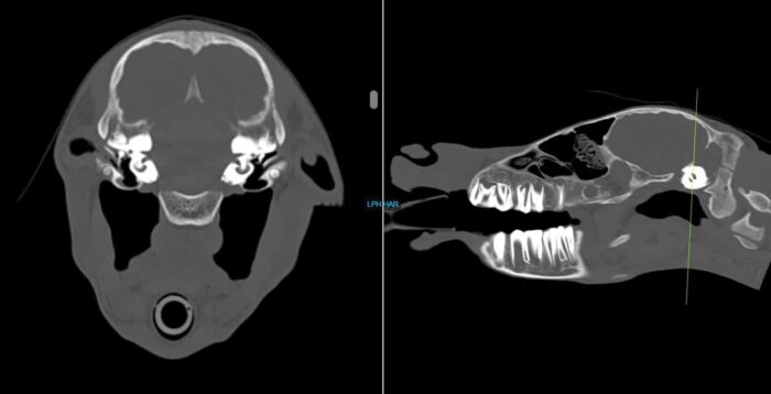
William is a 15 year old Warmblood gelding with a recent history of right sided facial swelling. William was referred to Animal Imaging for the use of the standing CT to evaluate the skull in fine detail to search for a cause of the swelling.

Equine Standing MRI

Samuel, 13-year-old Paint horse gelding, presented to Animal Imaging for magnetic resonance imaging (MRI). He has a +/- 3-year history of bilateral grade 3/5 forefeet lameness that improves 90% to diagnostic anesthesia of the abaxial sesamoidean nerves with suspect navicular or soft tissue injury based on previous radiographs and ultrasound.

CT of horse neck

Equine Head CT & MRI

Nova, an 8-day old Quarter Horse filly, presented to Animal Imaging following a 2-day history of right sided head tilt with no known evidence of trauma. She was worked up by a boarded internal medicine specialist who found no significant abnormalities on physical examination, bloodwork, and initial radiographic findings.