

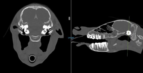
Nova, an 8-day old Quarter Horse filly, presented to Animal Imaging following a 2-day history of right sided head tilt with no known evidence of trauma. She was worked up by a boarded internal medicine specialist who found no significant abnormalities on physical examination, bloodwork, and initial radiographic findings. The filly presented for head and neck computed tomography (CT) to look for a possible cause of the head tilt and initial scan findings were relatively unremarkable aside from the appearance of debris seen within the right middle ear (blue arrow). Due to the lack of significant findings and the owner’s goals of identifying any significant performance limiting abnormalities within the foal, she was then taken to magnetic resonance imaging (MRI) for same day brain imaging. The collected images are shown below:
These images show cross sectional images of the skull of the foal. The image on the left shows a cross section taken from the area of the green line on the right-side image. As you can see with the blue arrow, the area of the right middle ear (right of the horse is on the left of the image) is grayer as compared with the clear area of black within the ear canal on the left of the horse. For this reason, MRI was elected to further evaluate this potential area of concern.
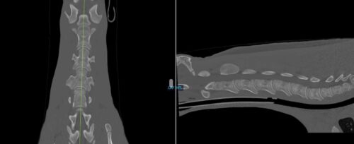
These images show a CT of the neck of a horse. The image on the left is a bird’s eye view of the vertebrae of the neck and the image on the right shows an image taken from the side of the horse’s neck.
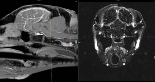
The following images are collected from the MRI of the brain of the foal. MRI collects images from multiple sequences to highlight different areas of normal brain tissue as well as potential pathology. The image on the left is taken as a cross section dividing the two hemispheres of the brain, and the image on the right is taken as a section shown through the green line on the image on the left. As you can see in the image on the right with the red arrow, there is an area of increased contrast within the cochlea of the right ear. This likely explains the foal’s clinical signs and the remaining brain appeared unremarkable.
Discussion
Through the use of advanced imaging, the veterinary team was able to assist the owner with overall decision making for the future of the foal. Both the collected CT and MRI studies were utilized to evaluate the structural conditions of the brain, skull, and neck of the foal. With all the information provided by the veterinary team, the owners can make the best decision for the treatment and potential future of the foal.