
Peach, a 11-year-old spayed female Golden Doodle, presented to her referring veterinarian for a mass that was palpated by the owner on the right elbow. The referring DVM was suspicious of soft tissue sarcoma and thoracic radiographs were collected at that time to evaluate for evidence of metastatic spread. On the radiographs, a 4cm right lung mass was seen. After discussion with the client, the owners desired additional information, including a complete chest evaluation with computed tomography (CT) as well as fine needle aspiration (FNA) to identify the type of cancerous lesions identified.
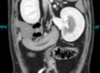
On presentation to Animal Imaging, Peach has normal vital parameters, behavior and normal bloodwork. A non-moveable mass was palpated overlying the right elbow region. She was placed under general anesthesia and a CT scan was performed, confirming the previously identified lung mass seen on radiographs. On the CT images shown below, we are able to clearly identify the large mass of the right lung, adjacent to the heart (seen by the blue arrow). These images shown here provide a transverse section on the left and a dorsal section on the right. In this manner with multiple sections of images collected, the entire 3D structure is able to be evaluated, therefore, being gold standard for checking for metastatic lesions, or spread of cancer in the chest.

One additional small nodule (blue arrows) was identified in the lungs with no additional areas of concern based on the study. These images below show the additional small area of potential concern. These images highlight the value that CT provides for identifying these more subtle changes in the lung tissue that otherwise might have been missed on radiographs until the mass was much larger. CT is able to remove the limitation of superimposition that radiographs are known for having when overlying a 3D structure on a 2D image.

Using 3D CT image guidance, a needle was guided directly into the larger of the two identified masses and sampled. The confirmatory image of correct needle placement is shown here.The patient recovered from anesthesia uneventfully and the collected tissue sample was submitted for cytologic diagnosis. After a few days, the results of the
submitted samples confirmed a diagnosis of a pulmonary carcinoma/adenocarcinoma.

Discussion
Using the information provided through a complete imaging and cytologic diagnosis, this patient was able to be referred to an oncologist for targeted therapy based on their specific diagnosis for both the disease processes as they were presented, i.e. treatment of both the elbow soft tissue sarcoma and the primary carcinoma found in the lungs.
This diagnosis was made possible with a team comfortable utilizing advanced imaging technology to provide the best quality of care for the referring veterinarians and the clients. Real time image guided procedures allow for the most accurate diagnosis available; these procedures minimize patient trauma, maximize anesthetic events, and provide safe, confirmed diagnoses for the patient care team. Customized, individual medicine allows for the client’s financial and emotional efforts to be directed intentionally at the confirmed diagnosis with minimal unnecessary treatments involved.
Please call to speak about additional cases of interest with the veterinary team here at Animal Imaging. We are skilled in making challenging diagnoses with minimal patient stress to help the veterinary team and the client have a safe and confident diagnostic experience.