Nuclear Medicine, also called Scintigraphy or Bone Scan is a form of imaging that uses small amounts of a radiopharmaceutical to diagnose or treat a variety of diseases. Animal Imaging provides nuclear medicine scans which include canine bone scans, thyroid scintigraphy, and trans-splenic portal scintigraphy.
Bone Scan
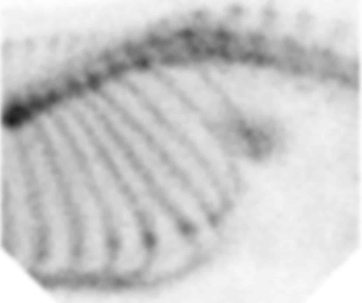
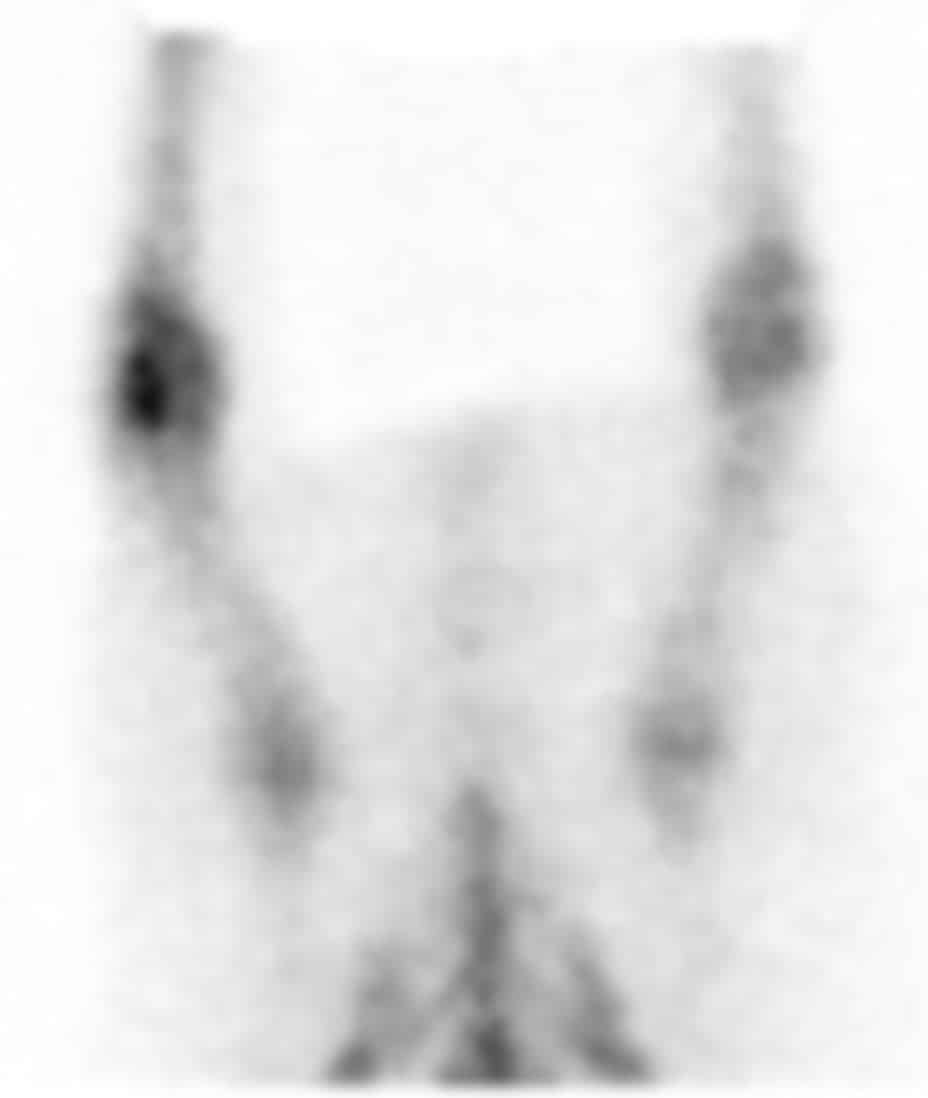
The Bone Scan (also known as scintigraphy) is a great resource for diagnosing obscure lameness issues. The patient is injected with a radioisotope which is then distributed throughout the body over a 2-hour period. The radioisotope that gets injected is absorbed in increased amounts in regions of the body undergoing remodeling processes. Using a special gamma camera, any areas of increased uptake can be identified and pursued further with other diagnostic techniques.
Common reasons for a Bone Scan
- Aid in the diagnosis of obscure lameness
- Aid in the detection of metastases in the axial and appendicular skeleton
- Diagnosis of occult or undiagnosed fractures
Trans-splenic Portal Scintigraphy for Portosystemic Shunts
Portosystemic shunts are abnormal vessels which allow portal blood to bypass the liver and enter the systemic circulation. Portosystemic shunts can be congenital (genetic) or acquired.
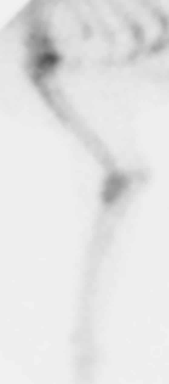
A diagnosis of portosystemic shunting is achieved at Animal Imaging with Trans-Splenic Portal Scintigraphy (also called Portal Scan). A small dose of radioisotope is administered under ultrasound guidance into the spleen. The passage of radioisotope is evaluated in real-time to determine if a macroscopic shunt is present and if the pattern of uptake supports a single congenital shunt or multiple acquired shunts.
If shunting is confirmed, this procedure is complemented with an abdominal CT scan to further evaluate the intra-abdominal organs and further characterize the shunting vessel(s). If no shunt is noted on scintigraphy, an abdominal ultrasound is performed for further information.
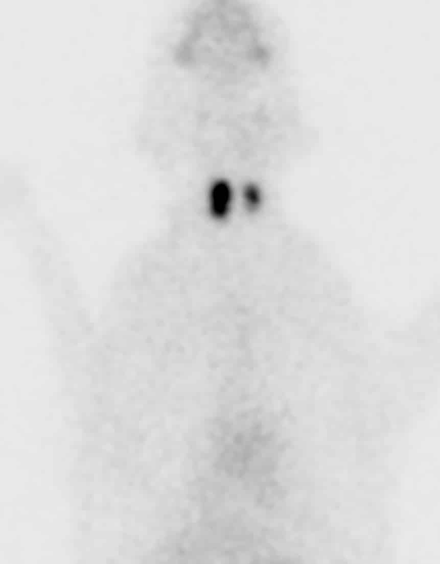
Thyroid Scan
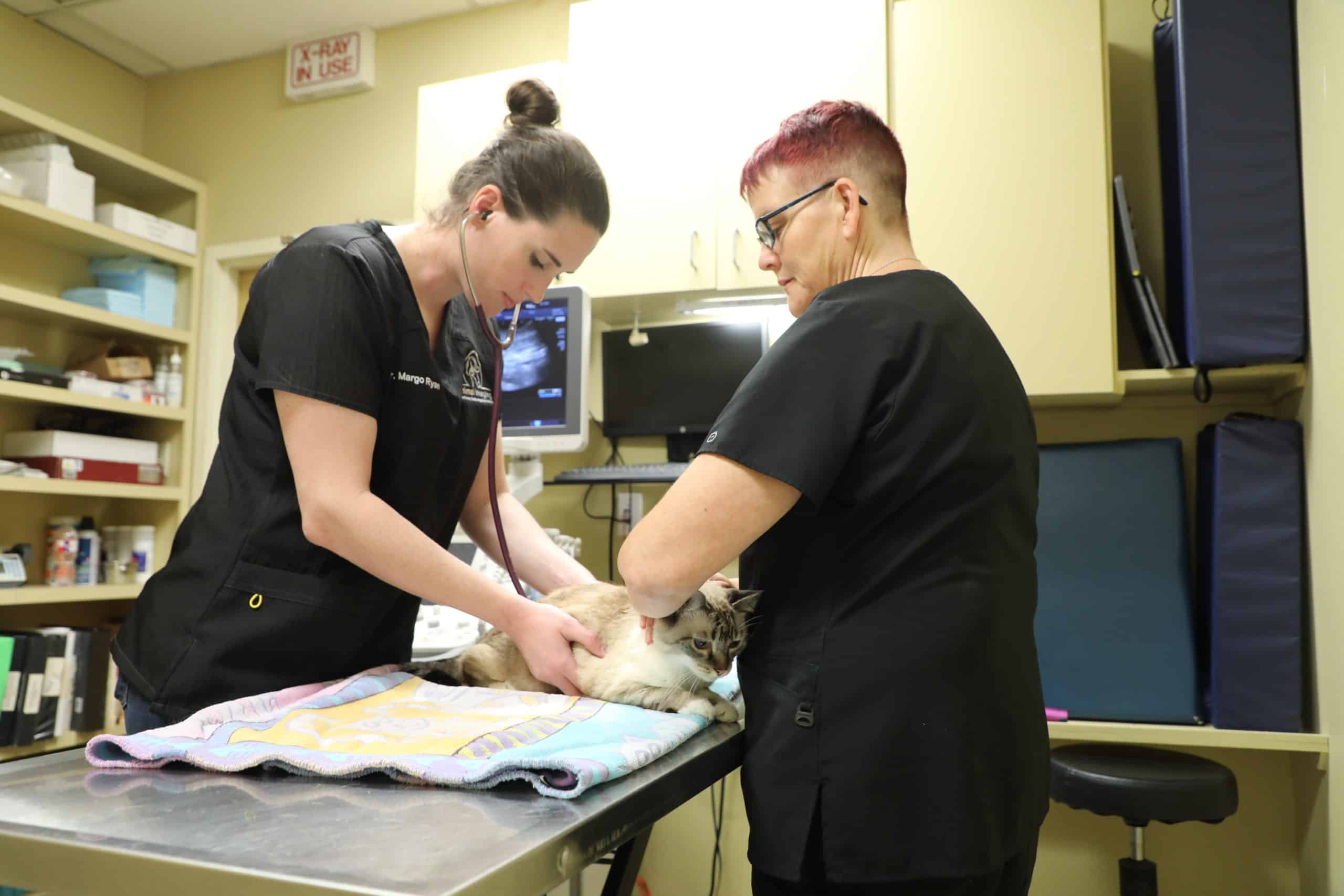
The Thyroid Scan is the first of a two-step process when a feline patient is being considered for Radioactive Iodine Treatment (I-131) to treat hyperthyroidism. This first appointment helps us determine, from a nuclear medicine standpoint, if the patient is a good candidate for treatment and to determine what dose of the I-131 would be needed to effectively treat the hyperthyroidism. This appointment is also used to discuss all risks, instructions, and concerns for the treatment process.
Radioactive iodine is an effective and safe treatment for hyperthyroidism. Patients are administered I-131 on Monday and hospitalized/boarded until discharge on Friday. Once home, the patient will need to be isolated for 2 weeks. More information can be found here.

What to Expect
Following the designated procedure, one of our board-certified radiologists will review all the images and submit a final report within 24-48 hours. The report and clinical findings will then be sent to the referring veterinarian. Clients should follow up with the referring veterinarian for explanation of results and information on treatment or management.
We are here to help.
At Animal Imaging, our passion is providing the best care and information to you and your veterinarian.
We are proud to have a dedicated team of board-certified veterinary radiologists available for your pet.
Questions or concerns? Call us!
(972) 869-2180
BE IN THE KNOW
Enter your email to be the first to hear about the latest case studies, announcements, and upcoming exclusive CE events.