
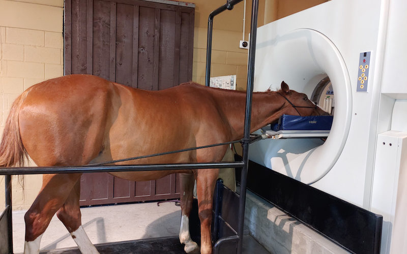


Cervical & Thoracic Spine Canine MRI
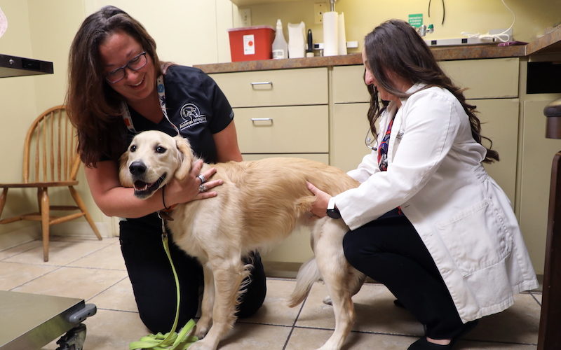
Marty, a 5 year old male neutered Labrador Retriever, was brought to the emergency room after Marty became painful and started to limp. There was no traumatic event the owners could recall and the examination at the emergency room found nothing of significance.


