
Benji, a 1-year-old intact male Golden Retriever, presented to Animal Imaging for advanced imaging of his right shoulder, brachial plexus, and elbow. He had a 9-month history of right front limb lameness that was worse after prolonged periods of laying down or intense exercise. He was moderately resistant to elbow extension on examination.
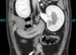
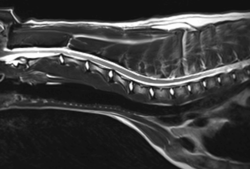
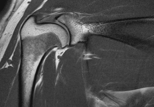
On presentation, Benji was grade 3/5 right forelimb lame. He was moderately resistant to right elbow extension. Shoulder radiographs were unremarkable, and elbow radiographs showed mild sclerosis of the ulna. He was anesthetized for magnetic resonance imaging (MRI) of the right shoulder and brachial plexus and bilateral shoulder and elbow/shoulder computed tomography (CT). The results of the images are shown below:
Diagnosis
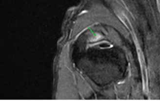
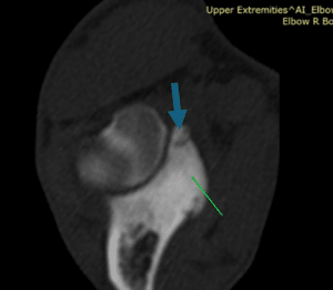
- Right elbow dysplasia characterized by medial coronoid process fragment.
- Moderate opposing stress sclerosis of the medial humeral condyle; no evidence of a subchondral bone defect.
- Secondary mild-moderate effusion and mild right elbow osteoarthritis
- Mild right supraspinatus tendinosis
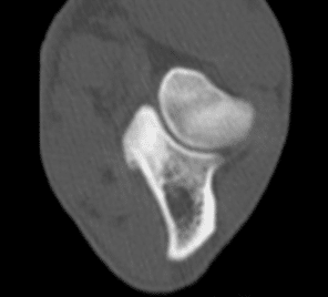
- Mild-moderate sclerosis of the left medial coronoid process; no fragmentation, likely stress sclerosis
Discussion
Due to the complementary nature of advanced imaging, both CT and MRI were used under one procedure to evaluate multiple potential causes of the patient’s lameness. The intervertebral discs, nerves of the brachial plexus, cartilage surface, soft tissue structures of the shoulders, and bone margins and internal architecture were all evaluated at a single time point. The use of concurrent advanced imaging allows the veterinarian to diagnosis and create a comprehensive treatment plan for the patient.
As you can see on the displayed images above, MRI allows for much clearer visualization of the soft tissue structures such as ligaments, tendons, nerves, and muscles as compared with the CT scan. The CT scan excels at providing clear information about bone detail and margins in a way that the MRI is not able to clearly delineate in the same manner. In addition, CT scans allow for rapid acquisition of images, thereby providing bilateral studies that allow for comparison images to be collected. Due to the nature of MRI physics, bilateral studies often are not in the interest of the patient, so lesion localization is performed prior to imaging to minimize anesthesia time and the associated risks. In this manner, the CT and MRI studies when used concurrently balance out the strengths and weaknesses of one another and allow for complete patient centric picture of their lameness to be identified.