
History
Copper is a 4 year female old Shih Tzu who had a history of elevated liver enzymes. Copper’s ALT was 700 (rr 0-120), pre bile acids were 84.7 (rr <13), and post bile acids were 256.4 (rr <25). With these lab values, the referring veterinarian was concerned for a possible shunt and Copper was referred to Animal Imaging.
Imaging
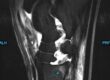
Initially, technetium 99 pertechnetate was injected in the splenic parenchyma under ultrasound guidance and dynamic scintigraphy was performed. The bolus of radioactivity coursed dorsally from the spleen and bypassed the liver into the thorax entering the heart followed by systemic circulation. This study confirmed the presence of a single congenital portosystemic shunt. From here, contrast-enhanced computed tomography was elected for further characterization and potential surgical planning.
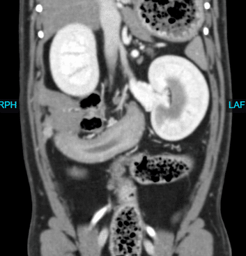
On CT, an approximately 6.2 mm in diameter tortuous vessel branched from the portal vein at the level of the pancreaticoduodenal vein and coursed caudally to enter the caudal vena cava immediately caudal to the porta hepatis, confirming the presence of a solitary extrahepatic portocaval shunt.
Another finding on the CT was a moderately sized, tortuous arterial vessels encompassing the left renal vein. A moderately sized (1 cm) wedge-shaped non-contrast enhancing region within the caudal pole of the left kidney was also present indicating an infarct.
Discussion
Copper had a clear diagnosis of a portosystemic shunt, as well as the surprising findings with the left kidney. Copper was diagnosed with left renal arteriovenous malformation with a moderately sized renal cortical infarction. Arteriovenous malformation is a vascular abnormality characterized by an abnormal tangle of arteries and veins which can lead to an increased risk of bleeding. They can occur anywhere throughout the body and can appear similar to vascular tumors such as hemangiosarcoma. The presence of nerve bundles differentiates arteriovenous malformation from tumors. These malformations can have a variety of treatments from conservative management to surgery where the lesion is either embolized or removed in cases of peripheral arteriovenous malformation.
Copper was also diagnosed with a portosystemic shunt, specifically an extrahepatic portocaval shunt. Normally, the portal vein brings deoxygenated blood from the abdomen into the liver carrying nutrients and waste products from the intestines, spleen, pancreas, and other organs. This blood then travels from the liver to the inferior vena cava to the heart. In Copper’s case, this blood is carried from the portal vein directly to the vena cava, bypassing the liver. This can cause symptoms like failure to thrive and abnormal behaviors. Bile acids are elevated in these dogs due to the release of the acids in response to feeding but not being reabsorbed by the liver. Treatment options can include supportive treatment with diet changes and medication, or surgical treatment to close the shunt and provides the best prognosis.