
Amir, a 21-year-old Arabian gelding, presented to Animal Imaging with a several year history of chronic, intermittent, purulent nasal discharge regardless of instituted therapy. He was managed with staged extractions of 108, 109, and 110 following dental radiographs and oral examinations. Post extraction radiographs are shown below.

The nasal discharge was persistent following dental extractions; therefore, antibiotic therapy and multiple sinus lavages were performed over the following months. The clinical signs remained, therefore surgical consultation for potential sinus flap and additional extractions was recommended by the primary veterinarian. To prognose dental and sinus disease, as well as offer surgical planning, computed tomography (CT) was performed by Animal Imaging. The corresponding CT images are displayed below.

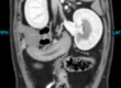
Equine Dorsal Skull section of the maxillary arcade. Blue arrows represent the108,109, and 110 alveoli with previously extracted teeth.

Inspissated material within the right rostral maxillary and ventral conchal sinuses. Small focus of mineral attenuation present.

Abnormal shape of the left first maxillary molar with mild widening of the periodontal space
Image Diagnoses:
- Moderate to marked right sinusitis, inspissated material within the right rostal maxillary and ventral conchal sinuses and a small mineral attenuating material in the rostral maxillary sinus
- Missing right fourth maxillary premolar (108) and right first and second maxillary molars (109-110) with likely oromaxillary fistulae in the empty alveolus of 110.
- Mild widening of the periodontal space of the left first maxillary molar and right third maxillary molar (inflammation vs early infection)
- Misshapen crown of the left first maxillary molar (previous fracture vs trauma)
Discussion
Computed tomography (CT) offers the gold standard for diagnosis of equine dental and sinus diseases. CT allows the veterinarians to thoroughly evaluate the entire periodontal ligament as it surrounds the tooth as well as identify areas of potential concern for close follow up examination on follow up oral and radiographic examinations. As shown in this case, CT allowed for a clear diagnosis of degree of sinus disease in a way that the radiographs were unable to. In addition, the CT imaging allows for surgical planning thereby decreasing time and associated trauma to animals. The identification crown fractures and periodontal widening on the contralateral side allowed for improved patient quality of care through close examination at the next time of procedure. This can decrease long term financial investments for the owners as well as decrease the number of sedated procedures through the animal’s life with earlier lesion identification.
Without the image challenges of soft tissue superposition, as seen with skull radiographs, CT allows for rapid, complete diagnosis of equine sinus and dental disease, ultimately improving quality of care and decreasing financial investments of the owners involved.