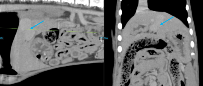
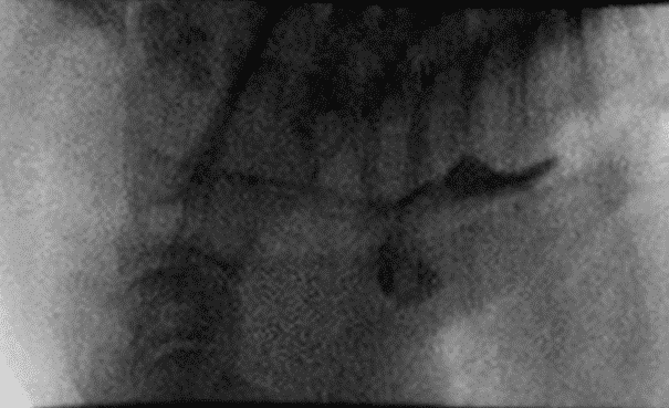
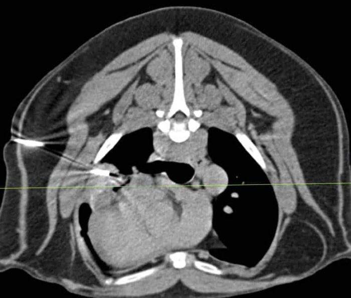
Canine Lung Mass FNA

Peach, a 11-year-old spayed female Golden Doodle, presented to her referring veterinarian for a mass that was palpated by the owner on the right elbow. The referring DVM was suspicious of soft tissue sarcoma and thoracic radiographs were collected at that time to evaluate for evidence of metastatic spread.
Equine Sinusitis

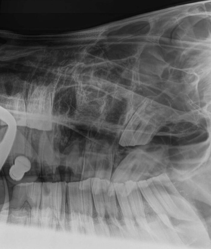
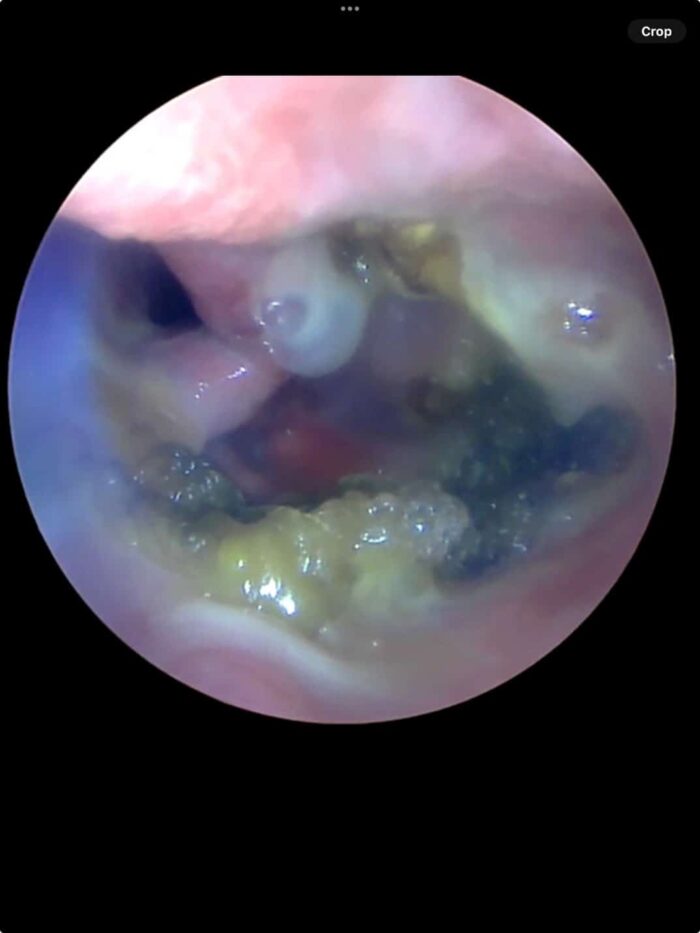
Majeed, a 21-year-old Arabian gelding, presented to Animal Imaging with a several year history of chronic, intermittent, purulent nasal discharge regardless of instituted therapy. He was managed with staged extractions of 108, 109, and 110 following dental radiographs and oral examinations. Post extraction radiographs are shown below.

Equine Standing MRI as a Recheck

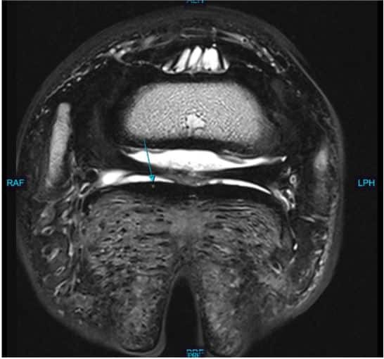
Storm, 9-year-old Holsteiner gelding used as a Jumper, presented to Animal Imaging for magnetic resonance imaging of the bilateral forefeet. He has an acute onset history of grade 2/5 right front limb lameness that resolves to diagnostic anesthesia of the right palmar digital nerves.

Equine MRI – Carpal Bone Fracture

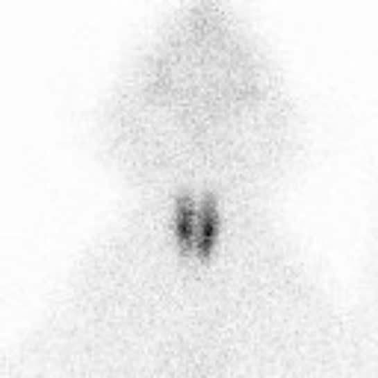
Dash, a 3-year-old Thoroughbred racehorse, presented to Animal Imaging for additional diagnostics after concern of decreased performance. A bilateral palmar digital nerve block improves the horse’s performance. Due to poor performance history, nuclear scintigraphy was performed to localize active sites of pain.