History
Wheatie is a 6-month-old intact female Vizsla who first presented to her referring veterinarian for recurrent urinary tract infections treated with repeated antibiotic therapy. As a board-certified internal medicine specialist, the veterinarian performed a complete diagnostic evaluation to identify a cause for the recurrent infections including bloodwork, urinalysis, abdominal radiographs, and an abdominal ultrasound. Here are the initial abdominal radiographs performed by the referring veterinarian:
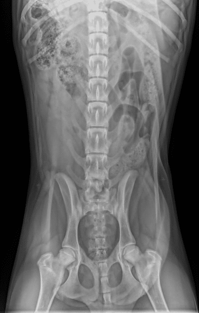
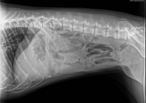
The abdominal radiographs are of excellent diagnostic quality, but no clear answer for the cause of chronic urinary tract infections is elucidated. Abdominal ultrasound is a relatively non-invasive modality that allows for detailed soft tissue evaluation, therefore is a logical next step in the diagnostic process.
The referring veterinarian then performed an abdominal ultrasound and identified an abnormal fluid filled structure to the right of the urinary bladder. Due to the abnormal anatomy visualized, the patient was referred for a second opinion and additional diagnostic imaging.
Upon presentation to Animal Imaging, a minor recessed vulva was appreciated on examination. An abdominal ultrasound was performed, and it was quickly observed that the urinary tract was abnormal. An enlarged left kidney, absent right kidney, and large fluid filled structure to the right of the urinary bladder was visualized. Two of the ultrasound images are shown here:
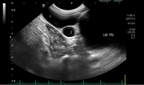
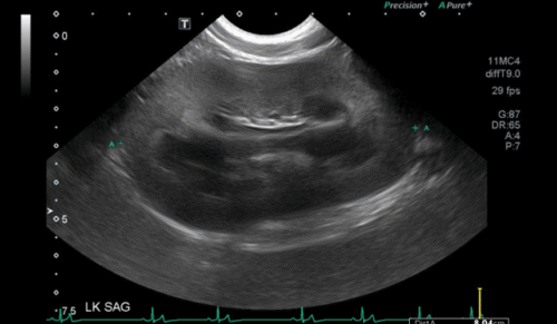
The abdominal ultrasound images shown here are representative of the multiple cystic structures (1&2) adjacent to the urinary bladder (UB TR) and the enlarged left kidney (LK).
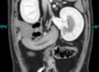
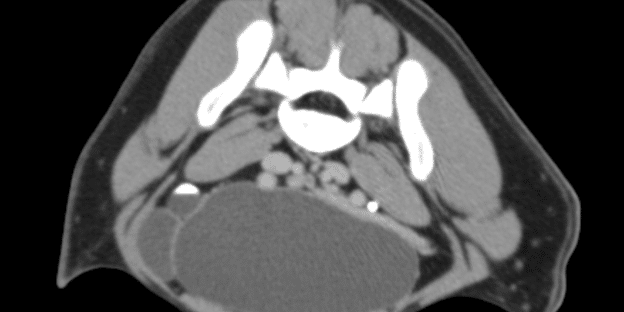
The owner was contacted and immediately informed that computed tomography (CT) scan would provide complete visualization of the abdominal organs, and the use of contrast would provide additional diagnostic information on the urinary tract patency. The use of contrast was important in this case given the lack of a right kidney visualized on abdominal ultrasound. The owner consented and an abdominal CT scan was performed. The CT images are shown here:
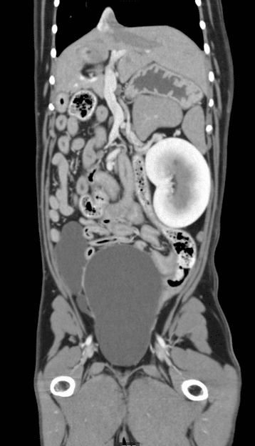
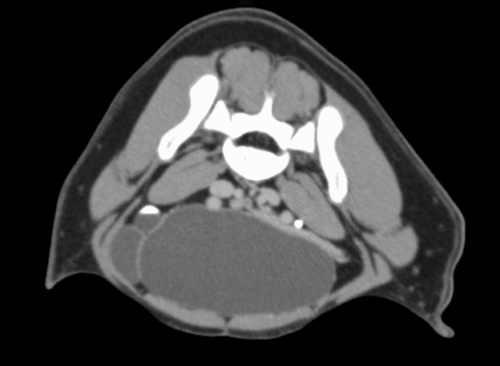
Multiplanar imaging as used in computed tomography allowed for diagnosis of the multiple fluid filled structures, as you can see on these images. Identification of any concurrent vascular, ureteral, renal, or other development abnormalities were able to be identified as well within the same abdominal CT scan.
The following conclusions were drawn from the acquired images on the case:
Image findings:
- Moderate to severe right uterine horn dilation with cystic remnant mesonephric or paramesonephric duct (e.g. Gartner cyst, Muellerian duct cyst) or congenital uterine anomaly such as uterine horn duplication with partial agenesis. Right uterine horn dilation may be secondary to pyometra, mucometra or hydrometra, and/or obstruction from congenital malformation such as stenosis.
- Right renal agenesis. Secondary left renal compensatory hypertrophy with small corticomedullary cyst. Mild left pyelectasia and mild hydroureter; physiologic or pyelonephritis/ureteritis.
The referring veterinarian was contacted with recommendations for an exploratory laparotomy and resection of the cystic structure with histopathology for a definitive diagnosis.
Discussion
This case shows an example of the value of multimodal imaging techniques as a means to complement the already completed diagnostics. The acquisition of excellent quality images allowed for a stepwise diagnostic plan to be implemented and ultimately reach the treatment recommendations in an efficient manner for the patient and client.
Abdominal radiographs were first utilized by the referring veterinarian to identify immediate reasons for the chronic, recurrent urinary tract infections, such as urinary stones. As you can see, superimposition of the gastrointestinal tract limits the soft tissue diagnostic ability of the cranial and dorsal abdomen on radiographs, regardless of the high-quality patient positioning and radiographic exposure technique. Both kidneys are difficult to clearly delineate, which is a relatively common occurrence, and not a cause for immediate concern or a clear answer to explain the recurrent urinary tract infections.
The images of the abdominal ultrasound show multiple cystic structures adjacent to the urinary bladder and a large left kidney. On occasion, additional abdominal structures, such as the colon, can create shadowing of the deeper abdominal structures that limits such visualization of normal anatomic structures. This is because any amount of gas in these structures, which is normal in a functioning gastrointestinal tract, is unable to be penetrated by the ultrasound beam, therefore appears like a black shadow on ultrasound. Due to this known limitation of ultrasonography, the use of repeated ultrasounds on different days, probes, or patient positioning helps to support any abnormal findings. Skilled ultra sonographers use a multitude of image manipulation techniques to confirm their findings; however, this is a known limitation of this modality. In this case, with already abnormal abdominal organs seen repeatedly on ultrasound by multiple highly trained specialist, abdominal CT provided the next diagnostic step to confirm the suspicion and identify any additional abnormalities that may have been missed.
CT allowed for the rapid acquisition of complete abdominal anatomical detail to confirm that the right kidney was not present and multiple fluid filled structures were present adjacent to the urinary bladder. Angiography is the use of an injected intravenous contrast adjacent that when paired with CT can be used to detail vasculature and the urinary tract. When used in cases such as this one, the use of contrast allows for the veterinary team to gain a sense of function and clinical significance of abnormal structures to advise the best treatment recommendations.
The use of multiple modalities allowed for complementary information to be gathered in a stepwise approach in this case. The high quality and timely manner for diagnostic imaging pursuits allowed the veterinary team was able to work effectively with the client to get an answer.