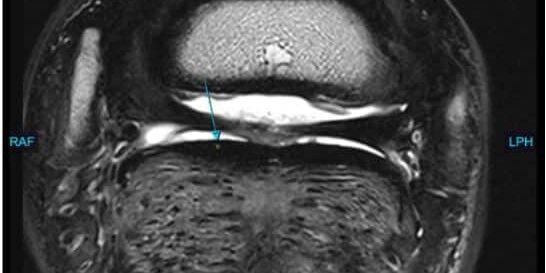
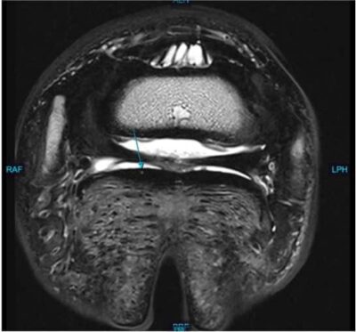
Storm, 9-year-old Holsteiner gelding used as a Jumper, presented to Animal Imaging for magnetic resonance imaging of the bilateral forefeet. He has an acute onset history of grade 2/5 right front limb lameness that resolves to diagnostic anesthesia of the right palmar digital nerves. He has repeatedly had both forelimb coffin joints and navicular bursas treated with steroid injections with a decreasing time of response to therapy upon repeated injections. Radiographs and ultrasound have been unremarkable with the primary veterinarian. He was first imaged by Animal Imaging with the 3 Tesla magnet (image left) which diagnosed a spiraling, pinpoint deep digital flexor tendon tear within the foot (blue arrow). Following treatment with biological therapy and rehabilitation, he was rechecked 9 months later via the standing Hallmarq 0.27T unit (image right) at the same level showing improvement of the deep digital flexor tendon lesion.
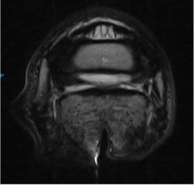
Another example is Brighton, a 7-year-old Quarter Horse mare used for barrel racing, who presented Animal Imaging for magnetic resonance imaging of the left hind limb. She had a grade 2/5 left hind limb lameness that was unchanged to flexion exam or diagnostic anesthesia. The left hind limb lameness was challenging to localize to one region of concern, so the proximal suspensory, fetlock and foot were imaged with the 3T magnet to identify the most likely pathology to explain the lameness. On initial imaging, the left hind proximal suspensory region showed mild changes, the fetlock was largely unremarkable, and a hoof defect was identified of the most dorsal aspect of the medial toe (image left). Following a period of rest for 3 months, the horse was reimaged using a standing MRI to see if the lesion identified within the hoof wall had changed. As you can see on the second provided image, the defect in the hoof wall and surrounding lamina was largely unchanged, increasing the suspicion of a likely keratoma in this case and therefore surgical consultation was pursued.
Discussion
The benefits of the 3T MRI include a high-quality resolution of study that allows for early small, detailed changes in soft tissue structures to be identified, as you can see here. A small pinpoint lesion in a flexor tendon, as seen in this horse, may be much more difficult to visualize on an image with lower resolution. The concern with using lower resolution studies to identify potential sources for lameness largely revolves around what potentially could start as a mild change and progress to a larger lesion if an early diagnosis is not made. Also, multiple scans can be performed back-to-back without requiring repeats as the images are collected without the patient moving. General anesthesia for these studies allows the fastest method of image acquisition as the patient is not moving during the procedure, enhancing the level of detail and speed of image acquisition.
The benefits of the standing MRI include the ability to perform the scan under standing sedation. For concerns of possible limb fracture, respiratory disease, or other contraindications of general anesthesia, the standing MRI offers a safe solution with minimal risk to the patient. A limitation of the standing MRI is patient movement. Whenever a patient moves during the time it takes to generate an image, the scan must be repeated, prolonging the amount of sedation and time for the procedure to be completed. In this manner, longer, multi-site studies with a poorly localized region for the lameness are less beneficial to the patient and cost to client as compared with a study under general anesthesia. In addition, regions imaged higher up on the leg of the horse are more sensitive to motion, as the horse may subtly sway or rock more than a foot would while they are sedated. This is why many horses that need imaging of the carpal or tarsal regions are better suited for imaging under general anesthesia. The standing MRI is best suited for recheck cases of lameness where the area of interest is clearly identified and minor changes in tissue architecture can be compared with lesion appearance from previous studies. Knowing key areas to recheck allows for targeted regions to image, decreasing the amount of sedation and time taken for the study to be completed.
There are many benefits and considerations of both imaging modalities utilized in our equine patients. Selecting the best modality for the diagnostic question at hand improves client satisfaction and patient quality of care when the best results are achieved with the lowest financial investment and risk undertaken. In this manner, the entire team at Animal Imaging is available to consult on modality selection for any case with the goal of providing answers for our patients and clients.


