History
A 10 year old neutered male Domestic Shorthair cat was referred to Animal Imaging for a thyroid scintigraphy and radiographs prior to radioactive iodine therapy. The patient had a history of weight loss and vomiting. On routine bloodwork, the total T4 level was elevated at 12.7 μg/dL (normal 0.8-4.7 μg/dL). The remaining bloodwork, including a CBC and biochemical profile, and urinalysis were within normal limits. The rDVM started the patient on methimazole therapy; however, the owners were having a difficult time treating the patient.
Physical Exam Findings
On presentation, the patient was thin on physical examination. A thyroid nodule was palpated in the cervical region. No cardiac murmurs were ausculted.
Image Findings: Thyroid Scintigraphy
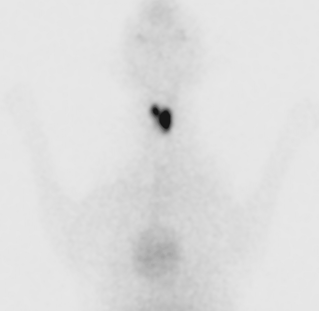
- Static images of the cervical region and thorax obtained after intravenous administration of a radionuclide.
- A smoothly marginated, ovoid region of increased radionuclide uptake is identified in the left mid cervical region, in the location of the left thyroid lobe. This uptake is increased relative to the ipsilateral zygomatic salivary gland with an elevated thyroid: salivary ratio of 6.6:1. A smaller focus of increased radionuclide uptake is identified in the right mid cervical region, in the location of the cranial right thyroid lobe. This uptake is also increased relative to the zygomatic salivary gland, also with an elevated thyroid: salivary ratio of 4.3:1. The remainder of the right thyroid lobe is not visualized, consistent with suppression.
Radiographs:
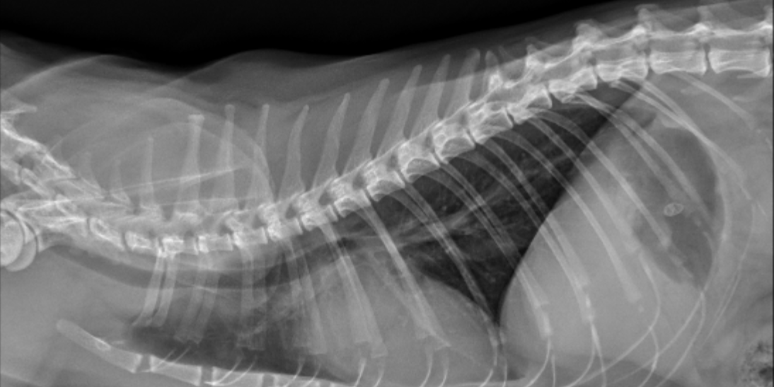
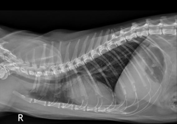
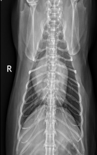
The cardiac silhouette is moderately generally enlarged. No abnormalities of the pulmonary vasculature or pulmonary parenchyma are identified.
Conclusion
Bilateral hyperthyroidism, worse on the left. The pattern of uptake is most suggestive of benign disease (i.e., adenomatous hyperplasia).
Moderate generalized cardiomegaly. The primary differences include: thyrotoxic cardiomyopathy or hypertrophic cardiomyopathy. No evidence of congestive heart failure.
Comments
Feline hyperthyroidism is a very common disease in older feline patients. Multiple treatment options, such as radioactive iodine (I-131), surgery, diet or medication (methimazole), are available and chosen based on the history, signalment, bloodwork findings, and comorbitities of the patient as well as treatment cost and availability. Radioactive iodine is often the preferred treatment choice among clinicians, but is only offered at approved locations, such as Animal Imaging.
Prior to treatment, thyroid scintigraphy and radiographs of the thorax and abdomen are performed. Thyroid scintigraphy is helpful if the disease is bilateral or unilateral and the pattern of uptake can be suggestive of benign versus malignant disease. This can be helpful to determine the best treatment option for a particular patient. Radiographs are performed to screen for concurrent disease, such as heart or kidney disease, as this may also influence the decision to treat with I-131.
In this case, I-131 was the preferred treatment option. The thyroid disease was bilateral making surgery (thyroidectomy) a poor choice. Oral medication (methimazole) was started but due to difficulty in medicating the cat, the owner did not want to continue with this treatment option. The patient did have concurrent heart disease but was clinically stable and deemed fit to undergo the treatment process.


