History
A two year old Quarter Horse mare was referred to Animal Imaging for a back-half bone scan. She initially presented with a grade 2/5 right hind lameness after suffering a fall in the wash rack. The right hind limb was blocked from the foot proximal to above the hock and there was no improvement of lameness. A back half bone scan was requested to help determine the source of the lameness.
Physical Exam Findings
Upon presentation the mare had physiological parameters within normal limits. She had some mild sensitivity to palpation of the right tarsus. The horse was very guarded to the manipulation of the lumbar vertebrae and pelvis.
Image Findings: Nuclear Scintigraphy
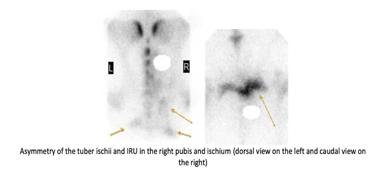
- Asymmetry of the tuber ischii with the right tuber ischium caudally located relative to the left and mild IRU in the region of the right tuber ischium, right ischial table, and right pubis. The primary differential is ischial and possible pubic fracture. A component of the IRU in the region may also be associated with artifact from urinary uptake, although this does not explain the asymmetric appearance of the ischii.
- Mild IRU in the region of the right coxofemoral joint/acetabulum as compared to the left. Differentials include fracture, osteoarthrosis, or artifact associated with positioning or overlying muscle atrophy.
Radiographs and Ultrasound:
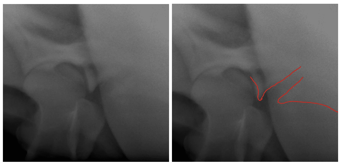
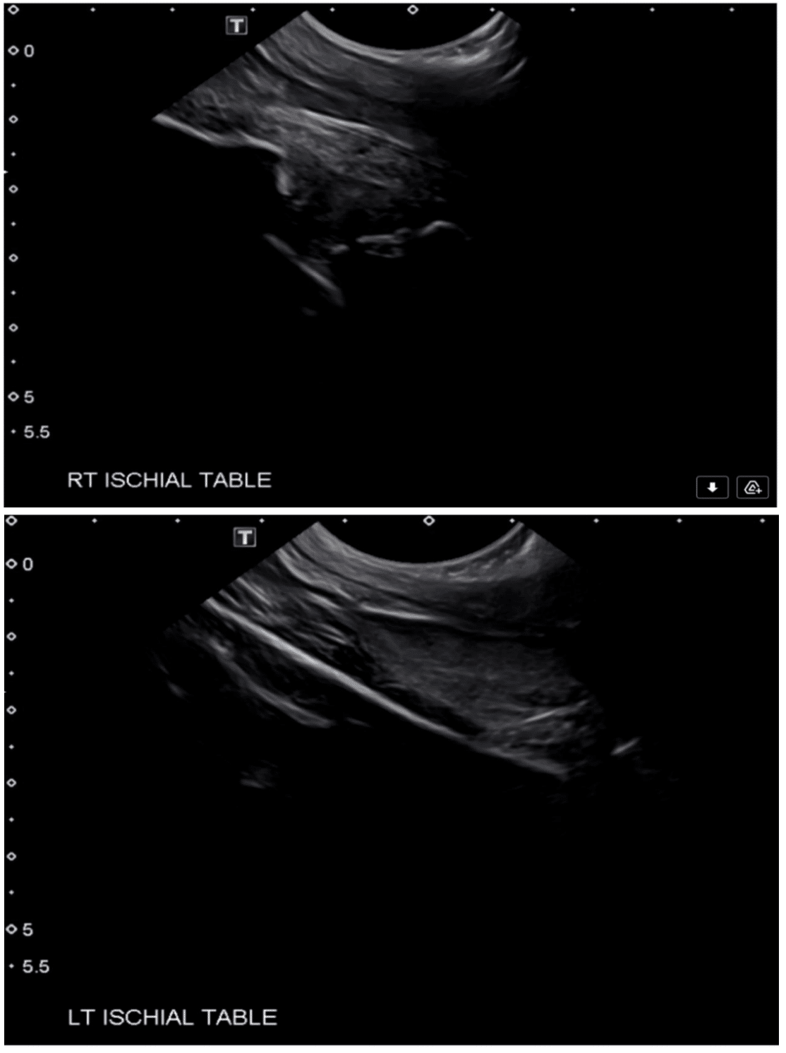
An obliqued, mildly caudally displaced fracture is identified in the right ischium immediately caudal to the acetabulum, visible radiographically and sonographically. There is no definitive communication to the acetabulum, although this possibility cannot be entirely excluded. An irregularly marginated step defect is also identified in the right ischial table transrectally.
Conclusion
Right ischial fracture, identified immediately caudal to the right acetabulum and extending along the ischial table. Although there is no definitive communication to the acetabulum, this possibility cannot be entirely excluded given the proximity of the fracture to the caudal aspect of the acetabulum.
Discrete fractures of the ischium are relatively uncommon. Ischial fractures that are articular (involving the acetabulum) and those that are displaced have a worse prognosis, with coxofemoral osteoarthritis affecting long term comfort in acetabular fractures and displacement affecting length of time needed for healing, as well as contributing to possible delayed or non-union. Rest should include at least one month of strict stall rest with the horse potentially being tied with its head up to prevent further trauma to the pelvis. After that period, another 2 months of stall rest is recommended followed by another 2 months of pasture rest. Possible slow return to work must be guided by assessment of bony union, soft tissue/muscle healing, and any persistent pain, keeping in mind the effect of an extended rest period on overall bone density. Repeat nuclear scintigraphy can be considered to help determine if there is still osseous turnover in the area.
References
Diagnosis and Management of Lameness in the Horse, 2nd Edition, Ross and Dyson


