History
Dancer, 3 year old spayed-female Sheepdog, presented to Animal Imaging for skull and thoracic computed tomography (CT) following a history of chronic epistaxis. On intake examination, the dog was mildly hypoproteinemic (5.1 g/dl) characterized by a hypoalbunemia (2.3 g/dL), and a mild inflammatory leukogram (neutrophillia 13,000/uL). All remaining CBC, ePOC, Chemistry, PT and PTT values were within normal limits.
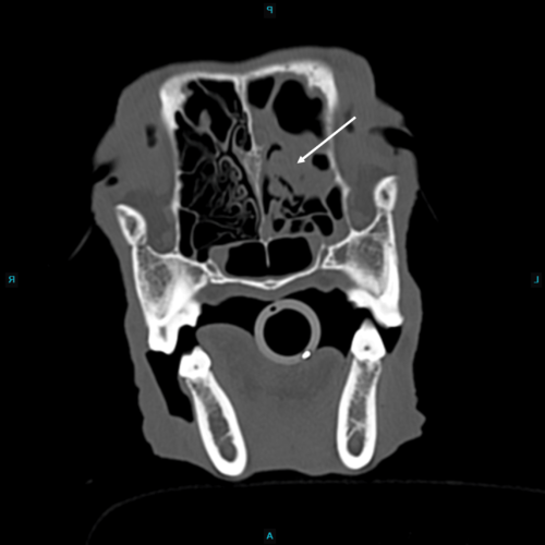
The results of the performed computed tomography were as follows:
1. Bilateral rhinitis, worse on the left, with bilateral nasal turbinate atrophy/lysis and left maxillary bone osteitis. (white arrow)
2. Moderate left frontal sinusitis with osteolysis of the frontal bone and left rostral dorsal calvarium, and osteitis of the left frontal bone.
3. Moderate to marked bilateral mandibular and mild left retropharyngeal lymphadenopathy. Differentials include: reactive/inflammatory change vs metastatic neoplasia (i.e., lymphoma).
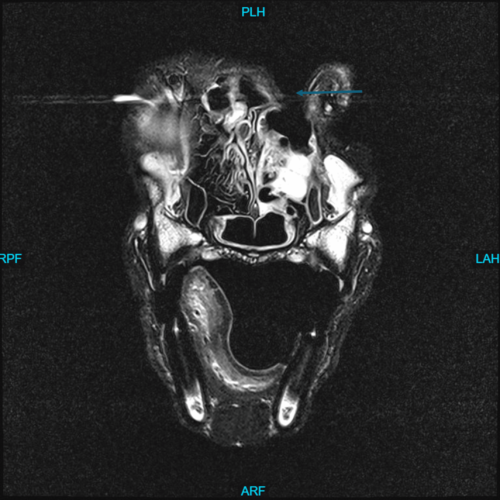
The differential list for these findings includes infectious (fungal) rhinitis, inflammatory rhinitis, or neoplasia (lymphoma). Rhinoscopy, nasal biopsy, and/or fungal titers were recommended, and the patient was treated for fungal rhinitis with clotrimazole by the referring DVM. He was responding to therapy for 6 months until he represented for acute grand mal seizure activity. Magnetic resonance imaging (MRI) was pursued to stage disease progression and the imaging results are shown below:
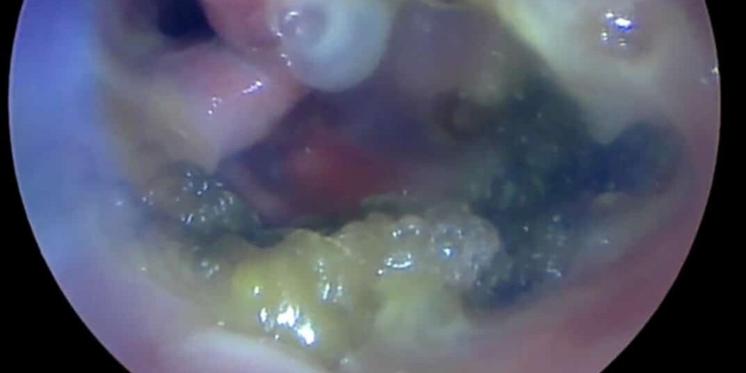
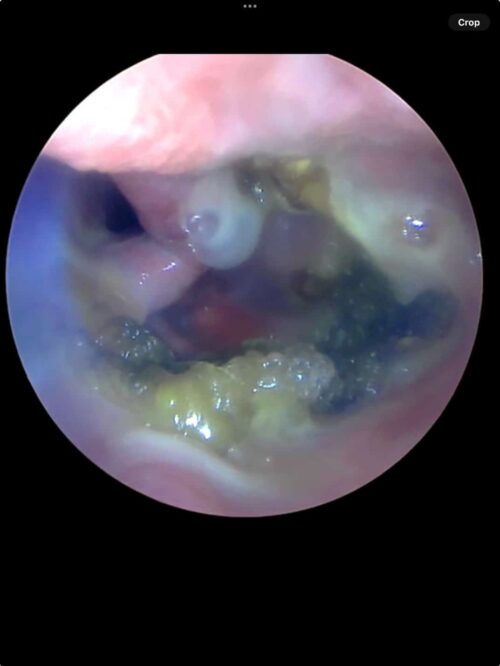
The results of MRI confirmed progressive, aggressive sinonasal disease with cutaneous fistula formation (blue arrow), mild intracranial disease, and regional meningitis consistent with Aspergillosis. Rhinoscopy was then completed for gross pathologic visualization while maximizing anesthetic events prior to waking the patient up. Rhinoscopy images showed a multifocal, necrotizing, lobulated lesion in the left sinonasal cavity. Gross visualization via endoscopy would allow biopsy when indicated.
In this way, multi-modal diagnostic imaging was utilized to repeatedly interpret clinical examination findings, stage disease progression, and offer prognostic information for improved patient care, diagnostic answers, and follow-up staging.
