History
Dash, a 3-year-old Thoroughbred racehorse, presented to Animal Imaging for additional diagnostics after concern of decreased performance. A bilateral palmar digital nerve block improves the horse’s performance. Due to poor performance history, nuclear scintigraphy was performed to localize active sites of pain. The imaging results showed:
- Left>>right third carpal subchondral bone injury, less likely osteoarthritis, unlikely adaptive remodeling
- Bilateral fore navicular contusion vs adaptive remodeling vs degeneration
- Bilateral dorsodistal P3 contusion vs adaptive remodeling vs pedal osteitis
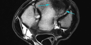
While the horse was still in the care of Animal Imaging, additional imaging of the carpi and distal fore limbs was performed to better qualify the source of increased uptake. Magnetic resonance imaging was performed, and the results showed:
Right Forefoot:
- Moderate asymmetric navicular bone edema like syndrome, likely secondary to inflammation, contusion or edema associated with trauma or aberrant loading
- Likely developing keratoma
- Mild generalized articular cartilage thinning of the distal interphalangeal joint
Left Forefoot:
- Moderate effusive navicular bursitis
Right Carpus:
- Maladaptive third carpal bone marked sclerotic remodeling and bone marrow lesion
- Increased vascular channels indicate increased abnormal bone stress
Left Carpus:
- Nondisplaced dorsal plane third carpal bone slab fracture
- Moderate-marked associated bone marrow lesion, likely contusion/inflammation
- Maladaptive sclerosis
To prepare for follow-up examinations, radiographs were taken of the left front carpus as well.
Discussion
Poor performance issues are a common presenting complaint when utilizing nuclear scintigraphy. This imaging modality examines large body regions and is beneficial in the interpretation of clinically relevant findings visualized on additional parts of the exam.
As seen in this case, this horse’s poor performance history led to a carpal bone fracture with no other clinical signs associated. As this boney surface is a non-weight bearing surface, these lesion types will impact performance but not ultimately cause lameness. The axial skeleton and the lumbopelvic regions are examples where nuclear scintigraphy assists with interpretation of clinical relevance in this manner.
In addition to nuclear scintigraphy, this horse received magnetic resonance imaging. Radiographs were offered as a diagnostic modality after nuclear scintigraphy, but the magnetic resonance imaging allowed for more specific findings to assist in prognostic grading and surgical assessment and planning. The use of advanced imaging modalities of nuclear scintigraphy and magnetic resonance imaging allowed for an accurate lesion diagnosis and therapeutic planning for the client and animal.


