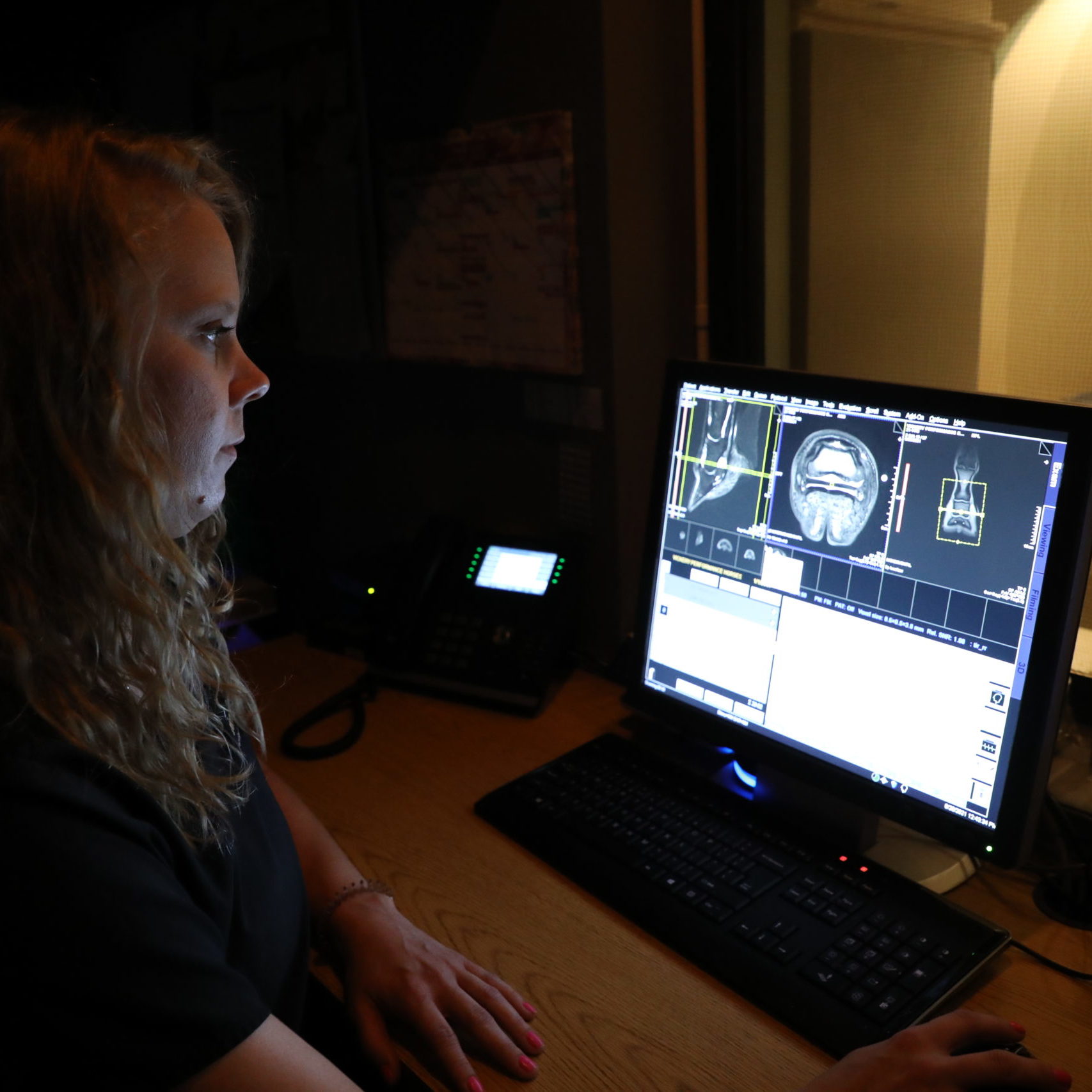
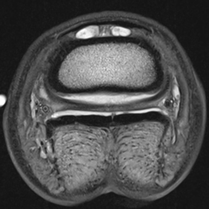
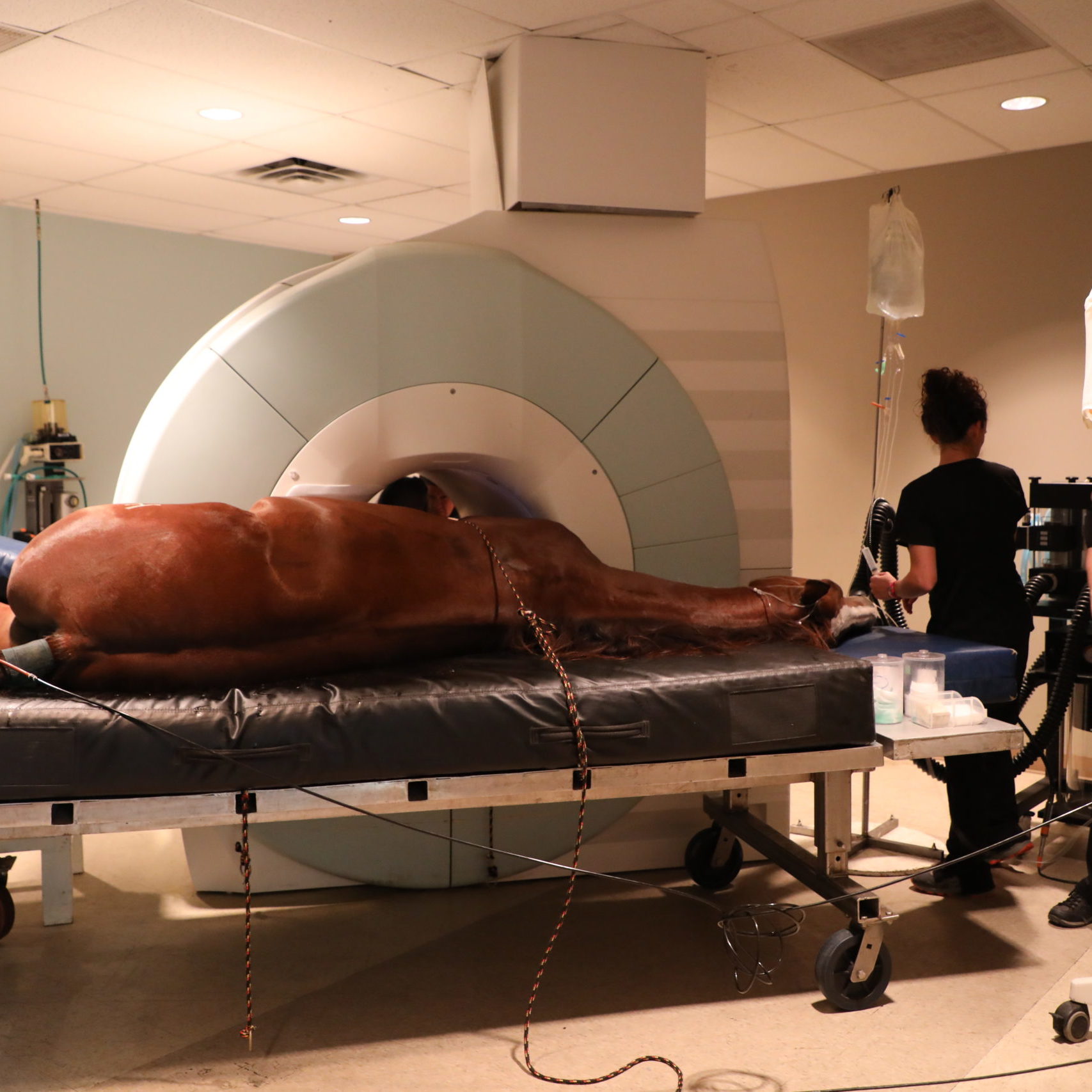
Magnetic Resonance Imaging (MRI) is a type of imaging that uses strong magnetic fields and radiowaves to produce detailed images of the tissues in the body. MRI is most commonly used for musculoskeletal injuries in the horse, especially in cases where radiographs and ultrasound are inconclusive.
Animal Imaging has the unique ability to offer two options for equine MRI examinations:
High Field MRI system: Animal Imaging is currently the only private practice in North Texas with a 3.0 Tesla MRI. The higher the Tesla strength, the faster the scan can be performed and the better the image quality. Due to the type and configuration of these high field systems, all patients must be placed under general anesthesia for the procedure.
Click here to learn more about 3T MRI from a recent Case Study.
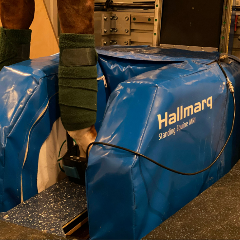
Low Field/Standing MRI System: Animal Imaging also has a 0.2 Tesla Hallmarq MRI on site. This system has the advantage of allowing the horses to remain standing during the procedure and avoid general anesthesia. The Standing MRI is used in addition to other services to help diagnose 90% of lameness. It is the gold standard in diagnostic imaging from the hoof to the carpus or tarsus.
Common reasons for an MRI at Animal Imaging include:
- Musculoskeletal Disease:
- Abnormalities of structures in the hoof
- Identifying pathology in soft tissue structures (i.e. ligaments, tendons) and bone.
- Assessment of many different synovial structures (i.e. bursa, joint, or tendon sheath).
- Neurologic disease localized to the brain
What to Expect
Upon arrival at our facility, please check in with the front desk. A staff member will discuss the procedure and answer any questions you may have. Prior to anesthesia, a veterinarian will perform a physical examination on your horse and remove any horseshoes as they must be pulled due to the properties of the magnet. A member of our team will advise you as to when your horse may be discharged dependent upon the type of MRI exam ordered by your veterinarian.
Following the MRI, one of our board-certified radiologists will review all the images and submit a final report within 24-48 hours of the exam. The report and clinical findings will then be sent to your referring veterinarian. Clients should follow-up with the referring veterinarian for explanation of results and information on treatment or management.
We are here to help.
At Animal Imaging, our passion is providing the best care and information to you and your veterinarian.
We are proud to have a dedicated team of board-certified veterinary radiologists available for your pet.
Questions or concerns? Call us!
(972) 869-2180
BE IN THE KNOW
Enter your email to be the first to hear about the latest case studies, announcements, and upcoming exclusive CE events.
