
History
Socks, 10-year-old male neutered Shepard mix, presented to Animal Imaging for abdominal ultrasound. He originally presented to his primary DVM after concerns of weight gain, polyuria and polydipsia, and previous diagnosis of hyperadrenocorticism. He was referred for detailed abdominal ultrasound to explain his now poorly controlled hyperadrenocorticism.
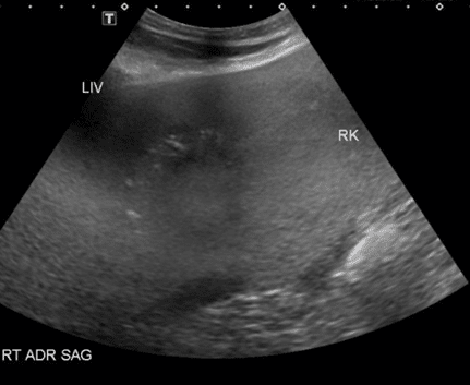
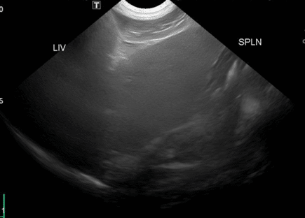
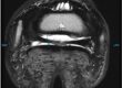
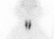
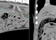
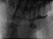
The first image shows a sagittal section of the right kidney and adrenal gland. Off the cranial pole of the right kidney, an enlarged, mixed echogenicity rounded structure of the adrenal gland is visible. In the second image, the spleen should normally be hyperechoic to the liver and in this patient the two organs are similar in echogenicity. The presence of the hyperechoic liver could represent steroid hepatopathy due to previous diagnosis of hyperadrenocorticism. An additional differential is hepatocellular carcinoma.
While still under light sedation for the procedure, a fine needle aspirate was able to be performed to ultimately rule out a potential neoplastic disease process for the patient and offer prognostic and treatment options to the owner.
Discussion
Many endocrinopathies require additional diagnostics to fully understand additional disease sequalae and management considerations based on the stage of the disease. Referral for additional imaging allows for complete abdominal evaluation and real time veterinary specialist interpretation of complex case presentations. This minimizes stress to the patient and client as additional diagnostics can be pursued on site and under the same procedure.