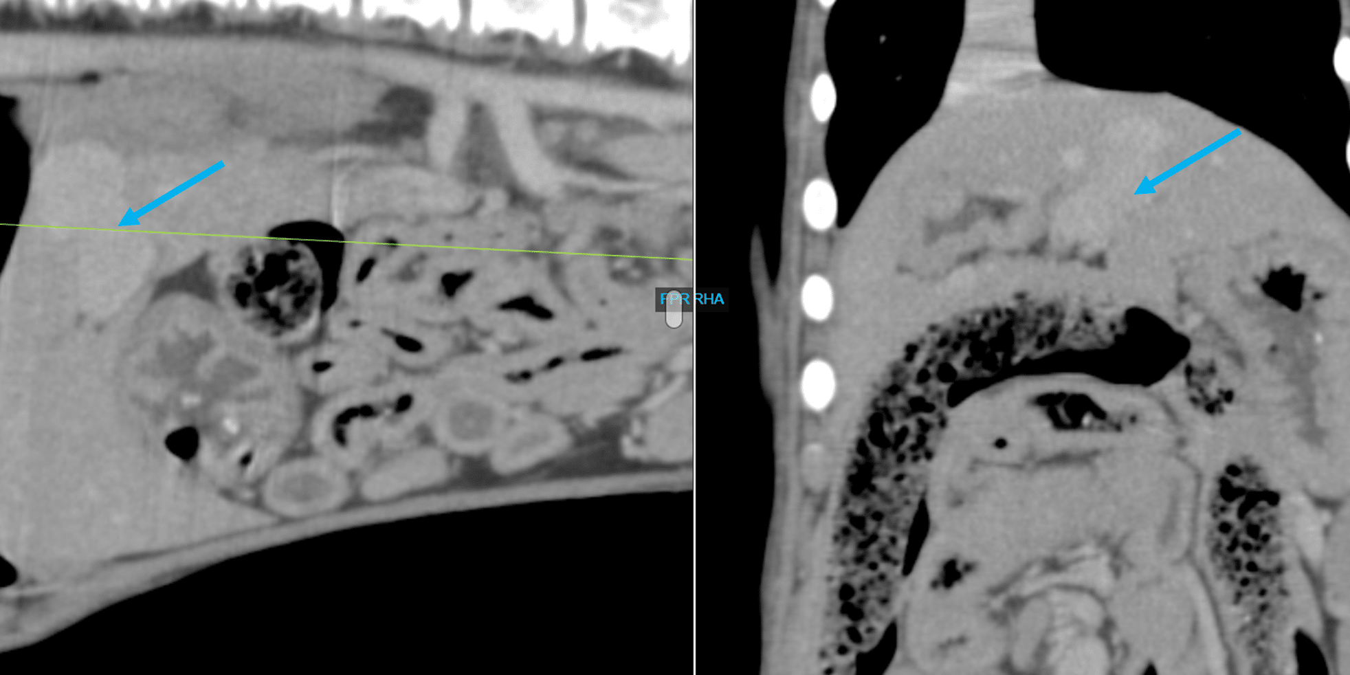
Kody, a 13-week-old male neutered Irish Wolfhound dog, presented to Animal Imaging for computed tomography (CT) after a history of being unthrifty with elevated bile acids and ALP on initial bloodwork. The abnormalities on initial bloodwork prompted the referral for computed tomography and angiography to look for the presence of a possible portosystemic shunt.
The resulting images from the CT scan are shown below:
Diagnosis
Intrahepatic portosystemic shunt.
Recommendations
Surgical ligation of aberrant vasculature
Discussion
Portosystemic shunts commonly present as a young-aged animal experiencing failure to thrive or ill thrift. Initial bloodwork shows elevations in ALP and bile acids leading to a referral for an abdominal CT for confirmational diagnosis and surgical planning. Broadly, portosystemic shunts are extrahepatic or intrahepatic with small breed dogs more commonly having of extrahepatic etiology compared with large breed dogs having intrahepatic shunts. CT angiography allows for tracking of the portal vasculature to visualize the presence of aberrant vasculature and surgical planning. This patient was returned to the referring DVM with recommendations for surgery. CT scans require a short procedure time, many requiring less than 30 minutes to complete, allowing for a short anesthesia and rapid report turnaround that offers veterinarians, clients, and patients quick answers to make additional plans for therapy. This patient was successfully treated by the primary DVM and able to return to his normal life after.


