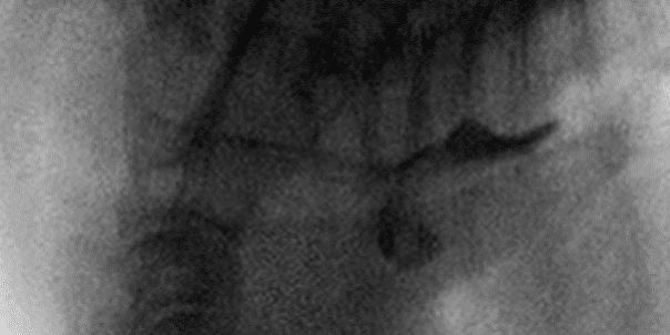
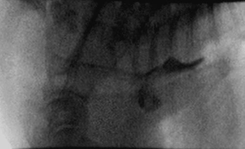
Tank, 4-year-old male neutered French Bulldog, presented to Animal Imaging for esophageal fluoroscopy after a history of chronic dysphagia and regurgitation. On presentation he had mild elevations in GGT (16 U/l) and T Bil (1.3 mg/dL) with no other significant findings. A small food bolus was covered in radio-opaque dye and he was allowed to eat the food while fluoroscopy camera was active to allow for visualization of the food bolus passing through the esophagus and into the stomach. The results of the imaging are shown below:
Discussion
The black radio-opaque material is highlighted coursing through the esophagus with the presence of a ventral deviation of food material present within the cranial thorax. The diagnosis for this case was a small cranial thoracic esophageal diverticulum with recommendations for medical management of esophageal dysmotility and possible esophageal endoscopy to allow for gross lesion visualization.
In this manner, dynamic fluoroscopy allowed for visualization of esophageal transit time, particularly as it relates to the presence of food material emptying vs remaining in the diagnosed esophageal diverticulum. Video images are provided to the reading radiologist that allow for complete prognostic grading of additional sequalae to the diverticulum including motility, gastroesophageal reflux, tracheal collapse, and laryngopharyngeal collapse. This modality allows for the real time interpretation of dynamic conditions that may be affecting many of these toy and brachycephalic breed type conditions. Dynamic imaging ability allows for information on disease conditions as it occurs in the patient’s life, thereby improving the lives of our patients and their caregiver’s ability to treat them as it uniquely relates to their lives.


