History
Gunner is an 8 year-old quarter horse used for barrel racing that was a grade 3/5 lame on the left hind limb with the lameness more evident when traveling to the right. No response to hoof testers. Negative to distal limb flexion despite palpable thickening of the soft tissues around the fetlock joint/suspensory branches. No improvement noted with plantar digital, abaxial sesamoidean, or low 6-point nerve anesthesia. Anesthesia of the proximal suspensory ligament improves the lameness approximately 85-90%. The tarsometatarsal, distal intertarsal, and tibiotarsal joints were treated with Arthramid. The proximal suspensory ligament was infiltrated with polysulfated glycosaminoglycan and betamethasone. Rehabilitation therapy was started. The horse failed to improve as expected with therapy and advanced imaging was recommended.
Initial examination matched with previous clinical examination. The horse received magnetic resonance imaging with ultrasonography performed by a boarded radiologist at the same time.
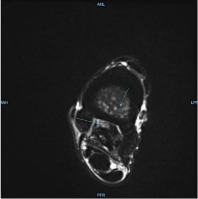
Findings on MRI included:
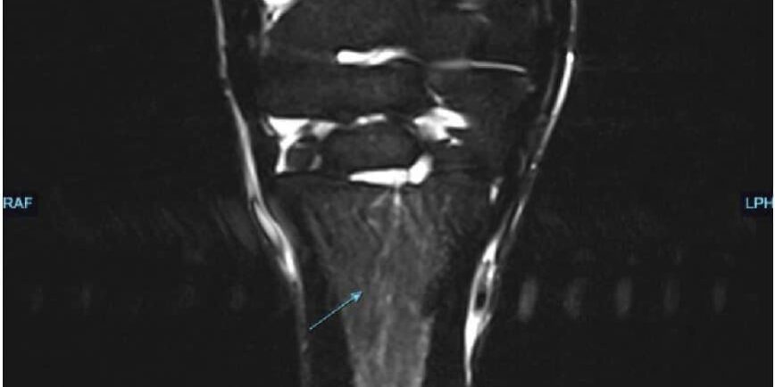
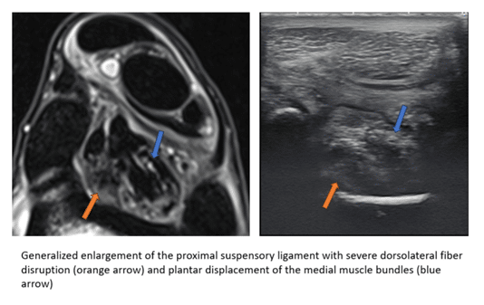
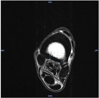
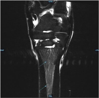
1. Severe proximal suspensory desmopathy and endosteal inflammation of MT3
2. Probable degenerative subchdondral lesion of T3/4 articulation. Clinical significance may be mild
3. Mild distal intertarsal and tarsometatarsal osteoarthropathy
Findings on ultrasonographic exam included:
The proximal suspensory ligament is markedly increased in cross-sectional area (2.35 cm²) with rounding of the plantar margin. There is severe dorsal fiber disruption resulting in loss of normal architecture and plantar displacement of the lateral and medial muscle bundles. These abnormalities extend approximately 4 cm distally. The plantar cortex of the third metatarsal bone is irregular with numerous punctate hyperechoic foci suggestive of mineralized foci or perhaps recent injectate.
Discussion
Magnetic resonance imaging allows the diagnosis of injuries not possible with other imaging modalities. Injuries are dynamic and necessitate follow-up examinations as well as follow-up imaging to further characterize changes/healing, as the initial MRI examination is simply characterizing the lesion at a single point in time. Monitoring lesions over time is essential for case management when determining workload and rehabilitation. Prevention of re-injury or worsening of the initial injury is paramount for successful case outcome. The timeframe for recheck imaging is dependent on initial lesion(s) and the clinical progression of the case. Thorough discussion with owners declaring the value of continued imaging as an essential component to rehabilitation and future soundness is important.
Follow up magnetic resonance imaging would be the best way to direct rehabilitation and monitor readiness for return to work in this case as both soft tissue lesions and bone edema were identified. Many clients may find follow-up MRI to be cost-prohibitive, which can limit the thoroughness of the rehabilitation period. In certain cases, recheck examinations in the standing MRI can be significantly more cost efficient as compared to initial imaging, although patient considerations as well as lesion considerations can limit this modality. For the client that has financial limitations, ultrasonography performed by a boarded radiologist at the same time as initial MRI offers the ability to have high quality recheck imaging at a significantly lower price than a second MRI. While most practitioners are well-versed in ultrasonography, there is considerable value in serial examinations being performed by a boarded radiologist that can compare to previous images (both US and MRI) without treatment bias. This case is an example of ultrasonographic imaging performed at the time of magnetic resonance imaging to establish baseline images for the purpose of recheck examinations to direct therapy and rehabilitation. Ultrasound was chosen for a more cost-effective way to follow the suspensory ligament lesion over time and help the referring veterinarian better direct therapy.
Dr. Meghann Lustgarten, who is boarded in both radiology (equine diagnostic imaging) and sports medicine, will be here monthly to assist with cases like this, provide follow-up imaging, and to meet the advanced ultrasound needs of our referring veterinarians.
