History
Orville is a 6 year old, male, neutered boxer that presented to Animal Imaging with a history of a left forelimb lameness of 3 weeks duration. Recent radiographs of the forelimb (shoulder, elbow, carpus, digits) were unremarkable. Lameness appeared to be progressing and on recheck examination, the cause of lameness could not be identified. Referral to an orthopedic surgeon was made and an appointment was obtained within the week of referral. On examination by the specialist, Orville was deemed generally healthy and bright. He was found to have a moderate weight bearing lameness of the left forelimb. The rest of the orthopedic examination was within normal limits as was a neurological examination. The source of pain could not be identified. Referral to Animal Imaging for nuclear scintigraphy (aka ‘Bone Scan’) was made.
Image Findings
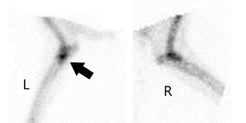
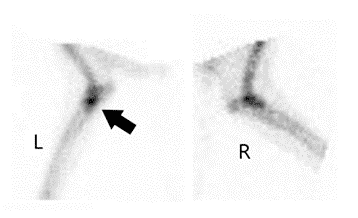
On nuclear scintigraphic examination, the most significant findings included moderate focal increased radiopharmaceutical uptake (IRU) of the medial coronoid process of the left fore ulna was noted. The primary differentials included: medial coronoid disease and/or a fracture/fissure of the medial coronoid process. Mild diffuse IRU was evident in bilateral elbows, the left shoulder, and the right carpus was identified. The primary differential for these joints was osteoarthrosis.
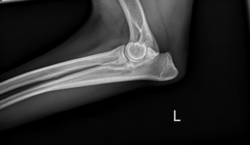
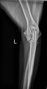
Radiographs of both elbows and the left shoulder were performed immediately following the scintigraphic examination. Medial coronoid disease of the left ulna was identified on radiographs (indistinction and irregularity of the medial coronoid process of the ulna on the lateral view with mild sclerosis along the distal trochlear notch of the ulna) with minimal/mild elbow osteoarthrosis bilaterally. Nuclear scintigraphy led to a definitive cause of the lameness.
Additional Imaging and Treatment Options
Further imaging such as computed tomography (CT) of the elbows could be performed to fully delineate changes in the joint that aren’t always present on radiographs due to superimposition. Targeted joint therapies like intraarticular injections of platelet-rich plasma or Synovetin OA® should be considered to improve pain/lameness without systemic effects.
Nuclear scintigraphy / bone scanning is a very useful tool in evaluating the skeletal system because actively metabolizing bone will have increased uptake of the radiopharmaceutical tracer, which allows identification of changes in bone metabolism before there is radiographic evidence of change. Bone scan in companion animals can be completed in several hours with the use of light sedation and animals can go home the same day as the procedure. Any condition which results in a change in the metabolism of the bone will result in a change in the appearance of the bone scan. Lesions like fractures, infections, tumors, and arthritis can be recognized with a bone scan long before they can be seen with plain radiographs.
One of the most useful applications for bone scintigraphy is in the evaluation of the patient with a poorly localized lameness. Localization of the source of a lameness in a companion animal is frequently difficult due to the inability of our patients to communicate and is complicated by the fact that many of our patients are extremely stoic and fail to react even when the painful area is directly manipulated. Many common causes for lameness in dogs and cats do not result in readily demonstrable radiographic lesions until the condition is advanced. This occurs because the structural or anatomic lesion may be extremely small. Fortunately, many of these lesions result in a significant change in the metabolism of the bone. This change in metabolism is what bone scintigraphy is able to detect.
By allowing the detection of the lesion early in the course of the disease, bone scintigraphy may allow for a surgical correction of the problem or medical intervention with disease-modifying therapies. Conditions causing lameness are usually best treated early in their course before the development of secondary arthritis which may be incurable. Joint arthroscopy and/or intra-articular therapy is best performed prior to significant radiographic changes, especially with disease-modifying therapy like platelet rich plasma (PRP) or pain-modifying therapy like Synovetin OA that prevents pain from becoming chronic. Tumors are obviously best treated early in the course of the disease before they have spread. Scintigraphy can identify boney tumors before radiographic changes are evident.


