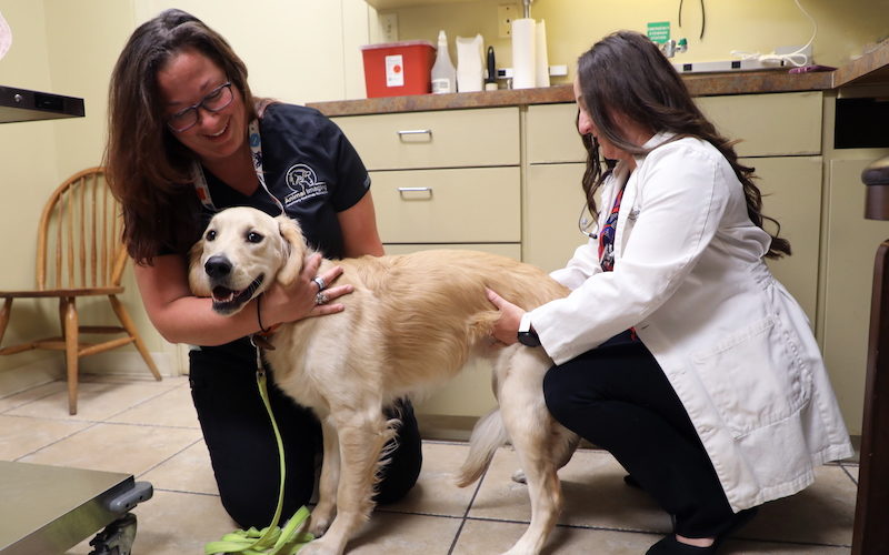
Equine Bone Scan
A two year old Quarter Horse mare was referred to Animal Imaging for a back-half bone scan.
Veterinary Radiology Specialists
Our team treats our patients as if they were their own pet and will ensure they are as comfortable as possible during their visit.
Utilizing some of the most advanced equipment and technology available, our board-certified doctors are able to see abnormalities, clarify findings, and provide a detailed report in a timely manner.
Being veterinarian owned allows us to stay unbiased in our commitment to providing the best specialty care available for our patients. Fast and accurate diagnostic imaging without bias.
At Animal Imaging, we value and care about our veterinarians and staff as much as we do for our patients and it shows in our collaborative and supportive work environment. Want to join our growing team?
A two year old Quarter Horse mare was referred to Animal Imaging for a back-half bone scan.
An eight year-old Quarter Horse gelding used for reined cow horse work, was referred to Animal Imaging for a 3T MRI of the left hind distal limb.
A 4-month-old female Pit Bull was referred to Animal Imaging for a (TSPS) to determine if the patient had a portosystemic shunt.
A nine year old, neutered male, labrador retriever with a history of 15 pound weight loss and intermittent GI upset was referred to Animal Imaging.
Enter your email to be the first to hear about the latest case studies, announcements, and upcoming exclusive CE events.